
Traumatic brain injury and mild traumatic brain injury (TBI and mTBI) are serious issues. Nearly 1.7 million traumatic brain injuries occur in the United States each year, according to the Centers for Disease Control and Prevention’s National Center for Injury and Prevention Control. Studies have shown that TBI accounts for over 50,000 deaths each year in the U.S. and over 70,000 patients with permanent neurological damage.
Diagnosing and properly treating TBI within the first 48-72 hours of injury can significantly alter the course of the injury and subsequent recovery. The quicker patients are properly diagnosed, the better, according to AuntMinnie.com, because after time, the ability to detect microbleeds falls, even though the chance of serious secondary injuries such as brain swelling or stroke is still there.
This has huge implications for the military, since 20-23 percent of military service members deployed to Afghanistan and Iraq have sustained a TBI while serving.
Use of MRI for early detection is helpful, but often unavailable for active duty military.
The results of a study of over 600 military personnel with TBI was published in Radiology on September 15, 2015. Authors Wei Liu, DSc, and colleagues used susceptibility-weighted magnetic resonance imaging (SWI-MRI) to monitor microhemorrhages in the subjects. The authors noted that 856 days is the average waiting period between the head injury and MRI scan for active duty military personnel who have sustained head trauma in the field; this is due to most brain injuries occurring in remote military action where there is no access to scanners. Researchers found that nearly a quarter of the patients had evidence of cerebral microhemorrhages within three months of their injury, but the number with detected bleeds fell in the months afterward.
Cerebral microhemorrhages can result from TBI and lead to severe secondary injuries, such as brain swelling or stroke, so the ability to detect them early is paramount. According to the study results, “the sooner an injured soldier received an MRI, the greater the chance of finding cerebral microbleeding.”
“The ability to monitor the evolution of microhemorrhages could provide important information regarding disease progression or recovery,” wrote lead author Wei Liu, DSc, and colleagues.
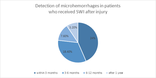
*Data from http://pubs.rsna.org/doi/full/10.1148/radiol.2015150160.
Children affected too
TBI affects people of all ages. In fact, according to a 2005 article by T. Babikian and colleagues, “traumatic brain injury is among the most frequent pediatric neurologic disorders in the United States, affecting multiple aspects of neuropsychologic functioning.” This study “assessed the efficacy of susceptibility weighted imaging as a predictor of long-term neuropsychologic functioning after pediatric brain injury compared with magnetic resonance spectroscopic imaging.” The abstract can be found on the National Center for Biotechnology Information website.
Final Thoughts | The Importance of Timely MRIs for Traumatic Brain Injury
Regardless of the patient’s age or occupation, the current research is compelling to seek MRI diagnostic testing as soon as reasonably possible when a suspected traumatic brain injury occurs. If you offer MRI services at your location, consider certifying your machine and your facility with RadSite accreditation.