
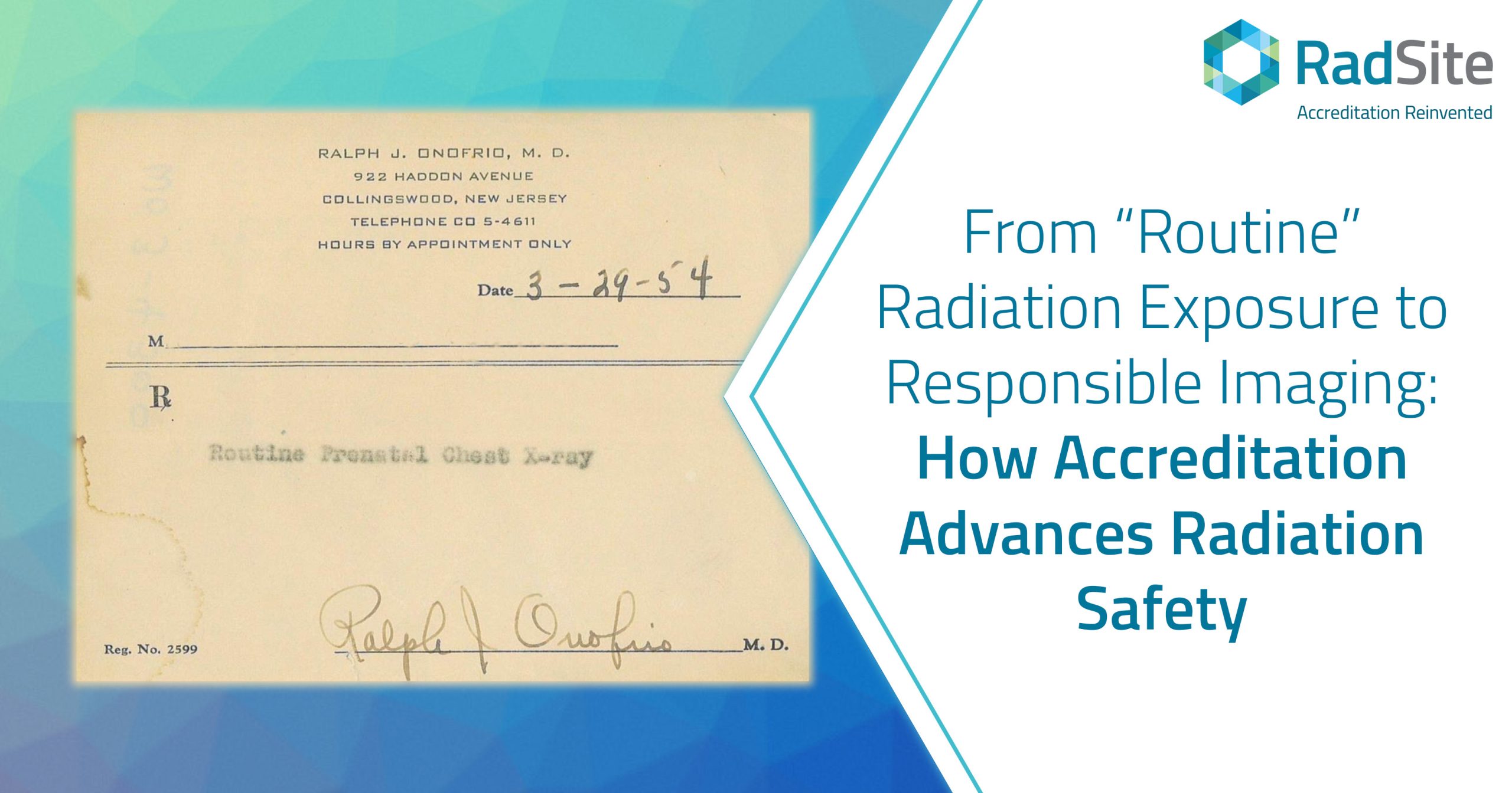
A framed prescription dated March 29, 1954, contains an instruction that now seems startling: "Routine Prenatal Chest X-ray."
The word routine may be the most striking part. The prescription captures a period when chest radiography could be incorporated into prenatal care as a routine examination, while the potential consequences of radiation exposure during pregnancy were less well understood.
Although RadSite does not accredit general radiography, the artifact offers a useful historical lens through which to consider how dramatically medical understanding—and medical common sense—can evolve.
At the time the prescription was written, the clinical benefits of X-ray technology were widely celebrated. The prescription illustrates how an imaging examination could become embedded in routine care before the patient-specific clinical justification and radiation-safety practices expected today were fully developed.
The artifact also belongs to a broader history of diagnostic radiation use during pregnancy. In 1956, British physician and epidemiologist Alice Stewart and her colleagues published a preliminary report describing an association between diagnostic irradiation in utero and childhood malignancy. A larger report followed in 1958. Their findings challenged prevailing assumptions and helped advance recognition of the potential risks associated with prenatal radiation exposure.
The lesson from that history is not that medical imaging should be avoided. Imaging has transformed diagnosis, treatment planning, and patient care. Rather, the lesson is that imaging should be undertaken deliberately: for a defined clinical purpose, using the safest appropriate modality and a protocol that balances diagnostic benefit with potential risk.
Radiation Safety Is a System
Radiation safety does not depend on a single policy, device, or individual. Instead, protecting patients requires a coordinated system encompassing clinical judgment, properly designed protocols, qualified personnel, equipment performance, quality control, medical physics oversight, documentation, and continuous improvement.
Accreditation helps assess whether these interconnected safeguards are present and functioning. By encouraging organizations to establish repeatable processes that can be evaluated, monitored, and improved, accreditation helps translate safety principles into practice.
RadSite's current accreditation portfolio includes CT, dental and medical cone-beam CT (CBCT), nuclear medicine and PET, MRI, and remote scanning services, with an ultrasound accreditation program under development.
Radiation-specific considerations apply most directly to modalities involving ionizing radiation, while other modalities and services have their own safety requirements. Across these programs, the common objective is high-quality imaging delivered within a disciplined patient-safety framework.
Safety Begins Before the Examination
The first radiation-safety question is not simply, "How much radiation will this examination deliver?" It is, "Will this examination provide information that is likely to improve the patient's evaluation or management?"
Accreditation does not replace the clinical judgment of the ordering professional or imaging provider. It can, however, reinforce an environment in which examinations are connected to defined clinical questions, available prior studies are considered, and protocols are selected intentionally rather than reflexively.
The goal is not to perform an examination out of habit or without a clear clinical purpose. Instead, it should provide information whose expected clinical value justifies its associated risks.
Optimization Does Not Mean the Lowest Possible Dose
Once an examination involving ionizing radiation is clinically warranted, the next responsibility is optimization.
Optimization does not mean reducing exposure so aggressively that the resulting images are no longer diagnostically useful. An inadequately performed examination can lead to uncertainty, delayed diagnosis, or repeat imaging—potentially exposing the patient to additional radiation without providing the intended benefit.
Instead, protocols should be tailored to the clinical task, the equipment, and relevant patient characteristics. The objective is to use radiation efficiently: enough to produce images of appropriate diagnostic quality, but no more than is reasonably necessary for that purpose.
This balance between radiation dose and image quality is particularly important in CT and CBCT, where acquisition parameters, field of view, positioning, and protocol selection can materially affect both exposure and diagnostic performance.
Physics Testing and Quality Control Matter
Even a well-designed protocol cannot compensate indefinitely for equipment that is improperly calibrated or whose performance has changed over time.
Routine quality control helps facilities detect changes in equipment performance. For imaging systems that use ionizing radiation, periodic evaluation by a qualified medical physicist provides a more comprehensive assessment of radiation output, image quality, and equipment performance.
Physics testing can identify subtle performance drift before it compromises patient care, results in repeat examinations, or contributes to unnecessary exposure. Such testing also helps verify that radiation output is accurate, reproducible, and appropriate for the clinical protocols being used.
Accreditation brings these activities together within a structured framework, helping ensure that testing is not merely performed but also reviewed and documented—and that identified deficiencies lead to appropriate corrective action.
Standardization Reduces Avoidable Variation
Variation is not inherently undesirable. Different patients and clinical questions may appropriately require different techniques. Unexplained or unmanaged variation, however, can signal inconsistency in protocols, training, equipment performance, or clinical workflow.
Standardization helps reduce unnecessary variation by establishing clear expectations for how imaging examinations are performed and evaluated. Consistent practices, in turn, support greater uniformity across facilities, equipment, and personnel.
This becomes increasingly important as imaging delivery models evolve. Remote scanning, mobile services, and increasingly sophisticated equipment can expand access and operational flexibility, but they also require clearly defined responsibilities, communication pathways, escalation procedures, and quality oversight.
Accreditation Supports Continuous Improvement
Accreditation should not be viewed as a one-time exercise or merely as a certificate placed on a wall. Its greater value lies in encouraging an ongoing cycle:
Establish expectations. Implement the process. Measure performance. Identify deficiencies. Correct them. Reassess.
That cycle is essential because medical evidence, technology, and clinical practice continue to evolve. What appears reasonable today may be refined tomorrow as better data become available.
The framed March 29, 1954, prescription for a "Routine Prenatal Chest X-ray" demonstrates that point vividly. It reflects how an imaging examination could become routine under the standards and assumptions of its time. As evidence and safety practices evolve, accreditation helps translate new understanding into safer care.
Imaging Deliberately
The guiding principle should be neither to avoid imaging nor to perform it reflexively. It should be to image deliberately.
That principle can be stated simply:
Image for a defined clinical purpose. Select the safest appropriate modality. Optimize the examination for the patient and clinical question. Confirm that the equipment performs as intended. Monitor performance and improve as evidence evolves.
Most patients will never know the names of the researchers, medical physicists, technologists, clinicians, standards committees, or accreditation reviewers whose work supports these safeguards. They nevertheless benefit from a system designed to turn lessons from the past into safer practice today.
That is one of the central purposes of meaningful imaging accreditation.
By Mark Hiatt, MD, MBA, MS
Chief Medical Officer, RadSite